

THE REAL-WORLD BOARDS: Question #2
There's only one right I.J.

Welcome to the Real-World Boards. As in the real world, there may be no “right” answer. If you have a suggestion for a new board question, please email it to boards@pulmccm.org. Thanks for participating!
A 64-year-old man is brought from a skilled nursing facility for lethargy. He is mildly hypotensive and in acute kidney injury with a creatinine of 3 mg/dL. He is treated empirically for sepsis with shock with 30 cc/kg crystalloid and broad-spectrum antibiotics IV. After a few hours he seems to be “perking up” (interacting, requiring only low-dose norepinephrine, and making some urine). The admitting physician’s opinion is that his predominant problem is volume depletion and that the renal failure will improve with aggressive volume resuscitation.
Overnight, his hypotension worsens to require norepinephrine up to 0.20 mcg/kg/min, although he appears otherwise unchanged. The covering physician places a central venous catheter in the right internal jugular vein and gives additional IV fluid boluses.
The next day, urine output stops, creatinine is 6 mg/dL and BUN is 110 mg/dL. The nephrologist recommends starting renal replacement therapy.
Right IJ CVCs complicate subsequent dialysis access in the ICU
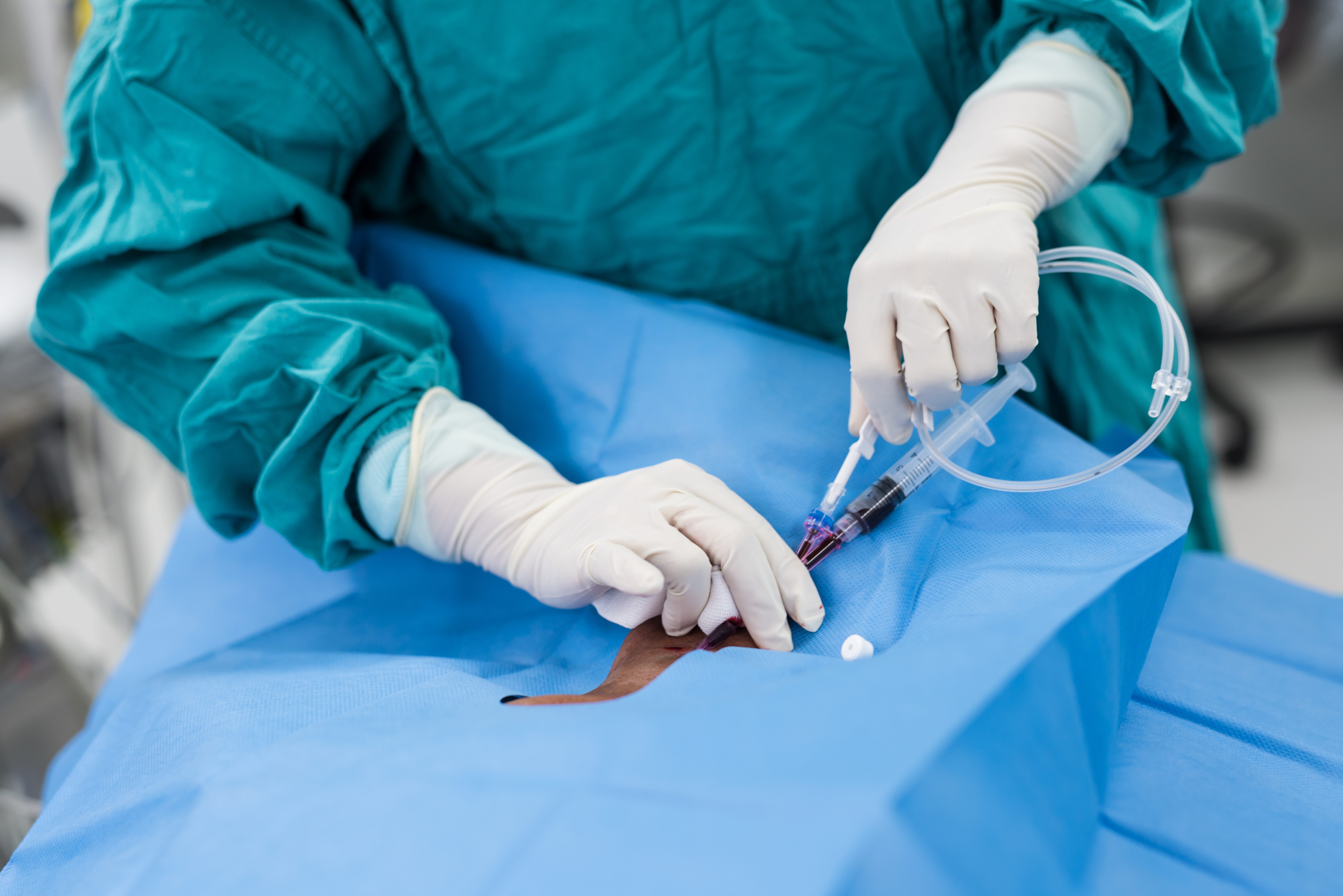
For patients who require temporary (or long-term) dialysis access, the right internal jugular vein site is preferred, due to the right vein’s larger diameter, straighter course, lower risk of stenosis/thrombosis, and better flow.
But ICU patients in shock or with poor venous access often require a central venous catheter before they develop severe acute kidney injury requiring dialysis. Most often, the right IJ vein is chosen for that central line, creating a challenge when dialysis access is later needed.
At the University of Maryland, 214 ICU patients requiring dialysis were retrospectively reviewed. About half had already had a central venous catheter placed in their left IJV (leaving the right IJ available for the dialysis catheter), and about half had one in their right IJ (requiring suboptimal dialysis catheter placement in the left IJV or femoral). (Gharaibeh et al, Journal of Critical Care 2025)
Patients with the right IJV already “blocked” by a CVC had much higher rates of later needing reinsertion of the dialysis catheter (40% vs 3% in the left IJ group).
They also required more central venous access procedures, with about 1 in 7 patients with an initial right IJ CVC requiring 4 or more procedures. Only 1 in 38 of patients with an initial left IJ CVC required 4 procedures.
Problems with left IJ dialysis catheters
To traverse the left IJ vein, the HD-CVC must make multiple turns before reaching the superior vena cava. Malpositioning with the need for reinsertion is common.
Tunneled left IJ catheters also seem to have a higher risk for infection, dysfunction, and migration, at least when they are positioned in the SVC. (Positioned deeper into the right atrium, they may function better.) Tunneled left IJ vein catheters may also have a higher association with central vein stenosis. Short-term left IJ dialysis catheters likely carry some stenosis risk, although data is lacking.
Right external jugular vein for dialysis catheters?
The right external jugular vein has been described in the interventional radiology and nephrology literature as a viable site for tunneled dialysis catheter placement when the right IJ vein is not available. However, this approach remains unfamiliar to most clinicians working in ICUs, and interventional radiologists will vary in their comfort level placing temporary vas-caths in the right EJ vein.
Right IJ vein, new stick vs. over-the-wire exchange
Removing the right CVC and placing a new dialysis catheter in the right IJ vein with a new stick might be considered the most conventional strategy. This may confer the lowest infection risk, but exposes the patient to the risks (pneumothorax, vascular injury, etc) of another procedure.
For patients in significant shock, it may be inadvisable to interrupt infusions of vasopressors or other vital medications for the duration of line removal and placement. If temporary peripheral IV access during the procedure is impossible, this strategy may therefore be risky or infeasible.
Exchanging a RIJ catheter over the wire is sometimes performed. This reduces the risk of pneumothorax, but is difficult to complete while adhering to strict aseptic technique, which is probably why higher rates of infection have been observed in some cohort studies for guidewire-exchange line placements.
The available data is limited, however, and other series have reported no increased risk of infection after guidewire exchange of uninfected central lines, when performed by interventional radiologists (who may be better able to ensure aseptic technique in their procedural suites, compared to the ICU). Another series reported by nephrologists at Duke showed no increased infection risk with nontunneled dialysis catheters placed by guidewire exchange, which was echoed by a post hoc analysis of the ELVIS randomized trial (for conventional CVCs).
The primary issue with guidewire exchanges is that the existing catheter is contaminated and can’t be handled without breaking aseptic conditions. If this strategy is attempted, the catheter and entire site should first be thoroughly scrubbed with multiple rounds of chlorhexidine, and a second operator wearing a sterile gown and gloves should assist in handling the contaminated catheter.
Placing a dialysis catheter and CVC side-by-side in the right IJ vein
The right internal jugular vein is generally large enough to accommodate a conventional CVC and a dialysis catheter side-by-side. This is rarely done, due to technical difficulty and perceived increased risks for vein thrombosis, catheter dysfunction, or other complications.
At one U.S. center, a single surgeon reported his 97 personal cases of side-by-side cannulation with both catheter types in the right IJ vein in ICU patients, and compared them with a cohort of 63 of his ICU patients in whom he placed a tunneled dialysis catheter only. Thrombosis, line infection, and line malfunctions occurred nominally more often in the patients with side-by-side lines, although non-significantly (1-2% for each complication vs. 0%). There were no pneumothoraces. (Spitzer et al, Journal of Vascular Access 2020)
Notably, though, a literature search reveals no other reports of this practice. By the odd logic of malpractice law, clinicians pursuing this option would by definition be practicing outside the standard of care and thus more vulnerable to liability claims in the event of a complication.
Femoral vein vas-cath placement
Placement of a dialysis catheter in the femoral vein eliminates the risk of pneumothorax, but not vascular injury or bleeding (which can be much more severe in the retroperitoneal space, although rates may be <0.5% in the ultrasound era). This approach also preserves multiple-lumen access in the right IJ vein if it is needed.
The risk for bloodstream infection is widely believed to be higher with femoral catheters, and this association has been reported multiply (Merrer et al JAMA 2001; Arvaniti et al, Crit Care Med 2017; Oliver et al, Kidney Intl 2000)
Femoral lines in obese patients (especially those with panniculi overlying the thighs) are believed to be at especially increased risk for infection.
This presumption was called into question by the 3SITES trial which showed that conventional CVCs in the femoral vein did not become infected more often than those at the IJ site. Roughly 30% of patients in 3SITES were obese.

In another randomized trial of 750 hospitalized patients in France requiring dialysis initiation, femoral CVCs were infected less often overall than jugular catheters (as measured by both colonization and documented bloodstream infections), albeit non-significantly (Pariente et al, JAMA 2008). Femoral catheters were much more commonly colonized at the time of removal in obese patients, however.
Patients in these trials likely received meticulous infection prevention practices that should not be expected to be consistently duplicated in most care settings.
Should this issue change practice?
How important is this issue? After all, most patients in the ICU with acute kidney injury never require renal replacement therapy. (Estimates vary, but it’s probably fewer than 20%.)
However, a higher proportion of those with vasopressor-dependent shock and AKI will eventually require RRT during their ICU stay.
Placing trialysis catheters on all patients with AKI would be excessive and unnecessary. Because trialysis catheters generally have one regular IV access point (lumen), this could also disadvantage patients who will need continuous infusions and medications via two or three IVs.
Left IJ vein CVCs are themselves susceptible to malpositioning (although whether this requires reinsertion is debatable).
Subclavian vein CVCs free up the right IJ vein for dialysis access, and bring a lower risk for bloodstream infection. Though subclavian lines carry a higher risk of pneumothorax, that complication can be immediately recognized and treated. More problematically, subclavian lines can cause central vein stenosis, permanently precluding future dialysis access in that arm (for the relatively small minority of patients who will require permanent long-term dialysis). The National Kidney Foundation’s KDOQI 2006 guideline update suggests that “subclavian vein catheterization should be avoided for temporary access in patients with kidney disease.”
Subclavian vein cannulation may be a fading skill in any event, as clinicians have increasingly shifted to the less technically challenging ultrasound-guided approaches to the internal jugular or femoral sites, and placing far fewer central lines in general.